VR and AR Revolutionizing Medical Imaging
Virtual reality (VR) and Augmented Reality (AR) are considered as the combination of VR hardware, artistic vision, and computer science. VR substitutes real world with a simulated one and the primary goal of AR is to make life simpler by integrating virtual information to immediate surroundings of the real world environment. It aids in enhancing your interaction with the real world. As we know AR/VR technologies have taken the entertainment and gaming industry by storm. Furthermore this technology has a potential to transform the medical or healthcare industry in several ways with applications in training, radiology, oncology etc.
AR technologies have integrated the real-world aspects with Magnetic resonance imaging (MRI) and Computerized Tomography (CT) scans data thereby helping radiologists to see under the skin of patients with ease for studying internal organs, muscles, and bones. AR technologies could bring about revolutionary improvements in healthcare by reducing medical errors and increasing accuracy during surgeries. Additionally, it would give a better understanding of complex medical problems between doctors and patients. AR could aid physicians in revealing the exact body region in order to make incisions and administering injections. VR technologies are acting as aid for physicians to prepare them for better surgeries, to elucidate diagnosis to patient and to provide trainings. Figure below is an illustration on how doctor use a VR tool to plan surgeries on newborns' missing pulmonary arteries.
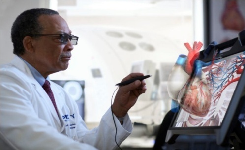
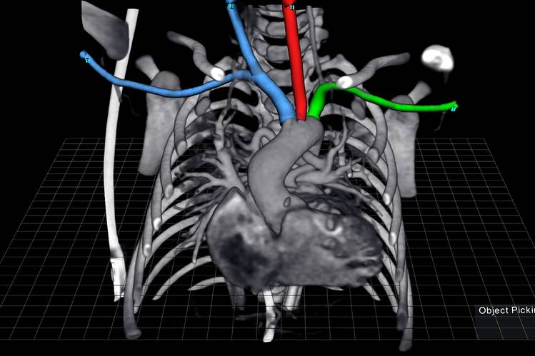
Image Source Ref.1
It is not an exaggeration to say that the integration of 3D images with VR will enhance the survival rate. In today's time most of the medical imaging are in the form of 2D images and physicians are required to utilize their skills & imagination to mentally sew together MRIs and CT scans in order to check how they fit together in 3D and quite understandably this task is very tedious and prone to errors. An AR system has been introduced that aids doctors to see real-time data from ultrasound probe directly onto a body of patient through an AR headset instead of a screen. This provides a single cohesive view rather than splitting the concentration of physicians between screen and patient. Physicians generally employed hand-held scanner to determine the blood vessels surrounding the wound. But the AR system helps them in locating those vessels directly by emphasizing them in 3D image as shown in an AR headset.
By: Dr. Deepika Koundal - Assistant Professor, CURIN
References
- www.stafordchildrens.org
- www.zdnet.com
CLICK HERE to Rate the Article |